A brittle star specimen was obtained subtidally at Oshoro Bay, Hokkaido, Japan, about 43°12′N, 140°51′E, on 30 June 2011 by Tadayuki Uramoto, photographed and fixed in 99% EtOH by Hiroshi Kajihara; the specimen was later identified as Amphiporis kochii Lücken, 1872 based on Kitagawa (2010). DNA was extracted from the disk of the animal using the silica method (Boom et al. 1990) with some modifications. Extracted DNA was dissolved in 30 ƒÊl of deionized water and has been preserved at ?20°C. Remaining morphological voucher specimen has been deposited at the Hokkaido University Museum under the catalogue number ICHU22080285 (contact: Hiroshi Kajihara, kazi@mail.sci.hokudai.ac.jp).
Amplification of mitochondrial cytochrome c oxidase subunit I gene (COI) using LCO1490 (5′-GGTCAACAAATCATAAAGATATTGG-3′) and HCO2198 (5′-TAAACTTCAGGGTGACCAAAAAATCA-3′) (Folmer et al. 1994) was unsuccessful.
An about 1000-bp fragment of 28S rRNA gene was amplified by polymerase chain reaction (PCR) using LSU5 (5′-ACCCGCTGAAYTTAAGCA-3′) andLSU3(5′-TCCTGAGGGAAACTTCGG-3′) (Littlewood et al. 1994). A hot start PCR was performed by a thermal cycler, iCycler (Bio-Rad), in a 20-µl reaction volume containing 1 µl of template total DNA (approximately 10–100 ng) and 19 µl of premix made with 632-µl deionized water, 80-µl Ex Taq Buffer (TaKara Bio), 64-µl dNTP (each 25 mM), 8-µl each primer (each 10 µM), and 0.1-µl TaKara Ex Taq (5 U/µl,TaKara Bio). Thermal cycling condition comprised an initial denaturation at 95°C for 30 sec; 30 cycles of denaturation at 95°C for 30 sec, annealing at 45°C for 30 sec, and elongation at 72°C for 45°C and a final elongation at 72°C for 7 min.
The PCR product was purified with the silica method (Boom et al. 1990). Both strands were sequenced with a BigDye® Terminator v3.1 Cycle Sequencing Kit (Applied Biosystems) following the manufacturer's protocol, using the same primer set as the initial PCR amplification. Sequencing was performed with ABI Prism 3730 DNA Analyzer (Applied Biosystems). Chromatogram and sequence data were operated with MEGA v4 software (Tamura et al. 2007).
Results
A total of 599 bp of 28S rDNA sequence was determined from Amphiphoris kochii (see Appendix).
Taxonomy
Phylum Echinodermata
Class Ophiuroidea
Order Ophiurida
Family Amphiuridae
Genus Amphipholis Thomas, 1966
Amphipholis kochii Lütken, 1872
(Fig. 1)
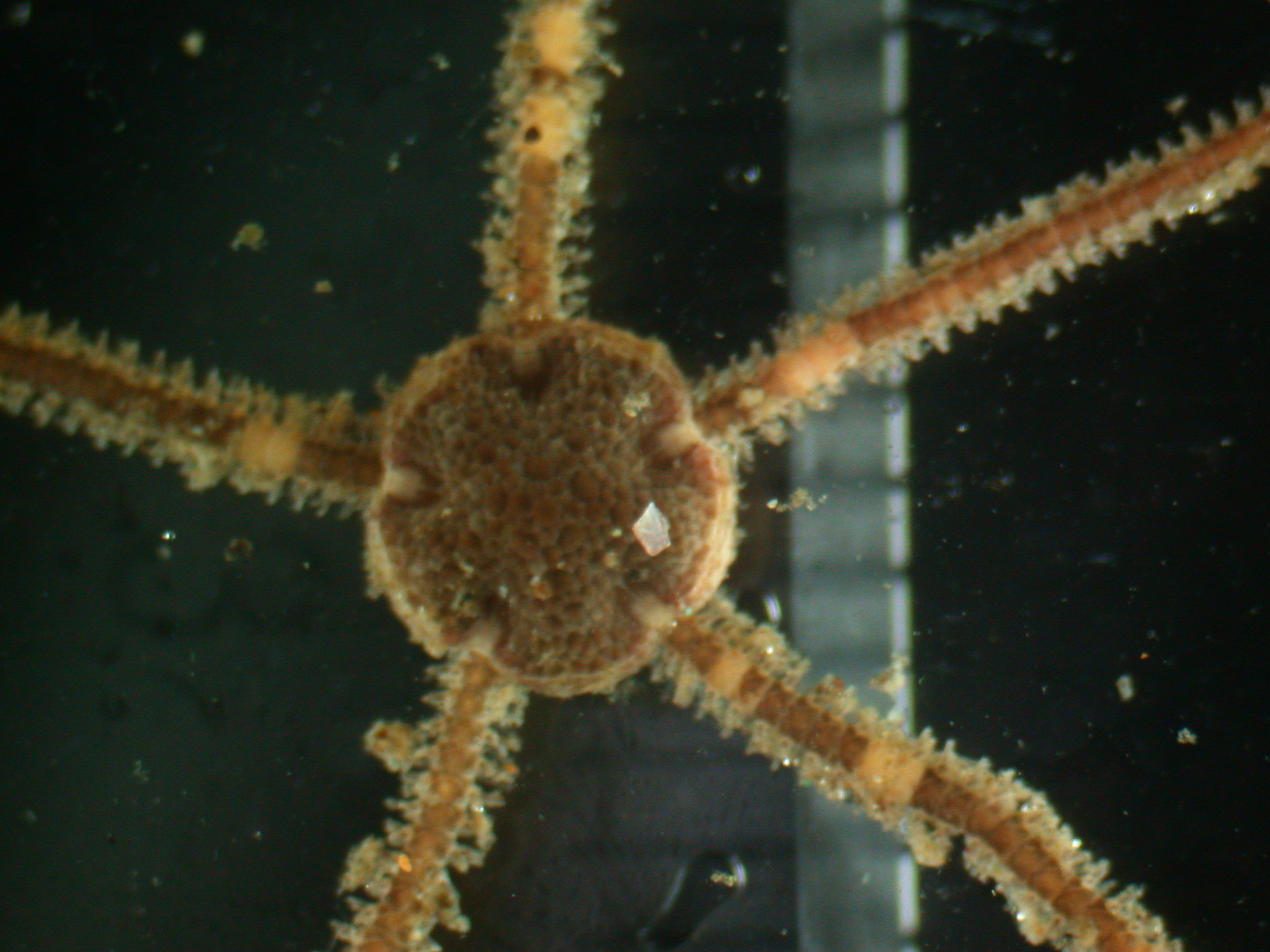
Fig. 1. Amphipholis kochii (ICHU22080285), aboral view.
References
Boom, R., Sol, C. J. A., Salimans, M. M. M., Jansen, C. L., Wertheim-van Dillen, P. M. E., and van der Noordaa, J. 1990. Rapid and simple method for purification of nucleic acids. Journal of Clinical Microbiology 28: 495–503.
Folmer, O., Black, M., Hoeh, W., Lutz, R. and Vrijenhoek, R. 1994. DNA primers for amplification of mitochondrial cytochrome c oxidase subunit I from diverse metazoan invertebrates. Molecular Marine Biology and Biotechnology 3: 294–299.
Littlewood, D. T. 1994. Molecular phylogenetics ofcupped oysters based on partial 28S rRNA gene sequences. Molecular Phylogenetics and Evolution 3: 221–229.
Tamura, K.,Dudley, J., Nei, M. and Kumar, S. 2007. MEGA4: Molecullar Evolutionary Genetics Analysis (MEGA) software version 4.0. Molecular Phylogenetics and Evolution 24: 1596–1599.
Appendix 28S rRNA gene sequence from ICHU22090063 identidied as Amphipholis kochii Lütken, 1872.
CGGTCTCGTGCCTAGTATTTAGCCTTAGATGGAGTTTACCACCTATCTTTGGGCTGCATTCCAAAACAACCCGACTCTTAGAAGCCTGATGGCCACCGGATACGGGAGCCTGTGGGGGCCTAACACACTCTATGGGATGAAGCCTCCTTCACAAGGACGTCGGCTCCACGTTTCCGATAAGCCACAGCTTCCTCACGCTACATTTCCCACACGCCGTTGAGGCGCGGGGATTTAGCGATGGGCTCTTCCCGCTTCACTCGCCGCTACT