A polychaete specimen was obtained from laminarian holdfast, which was collected at Oshoro Bay, Hokkaido, Japan (43°12′N, 140°51′E), on June 6, 2011, by Kentaro Miyashita. The picture of the specimen was saved, and then, identified by Hiroshi Kajihara to be a member of the family Syllidae. The specimen was fixed by soaking in 99% EtOH. DNA was extracted from the specimen using the silica method (Boom et al. 1990) with some modifications. A part of the fixed specimen was homogenized in a binding buffer. The homogenate was centrifuged at 12000 rpm. The supernatant obtained was put into a tube containing a 10 µl silica gel solution. After mixing them the solution was centrifuged at 12000 rpm. The supernatant was removed and a binding buffer (400 µl) was added to the residue. The solution was mixed again, and centrifuged as a same condition as described above. The supernatant was removed and a binding buffer (800 µl) was added to the residue, and then, DNA fraction bonded to the silica gel was recovered. This fraction was washed again with a binding buffer (800 µl). After drying in a air for 5–10 min extracted DNA was dissolved in 30 µl of deionized water and was preserved at –20°C. Remaining morphological voucher specimen was deposited at the Hokkaido University Museum under the catalogue number ICHU22090154 (contact: Dr. Hiroshi Kajihara, kazi@mail.sci.hokudai.ac.jp).
Amplification of an about 600-bp fragment of mitochondrial cytochrome c oxidase subunit I gene (COI) was attempted by polymerase chain reaction (PCR) using LCO1490 (5′-GGTCAACAAATCATAAAGATATTGG-3′) and HCO2198 (5′-TAAACTTCAGGGTGACCAAAAAATCA-3′) (Folmer et al. 1994). A hot start PCR was performed by a thermal cycler, iCycler (Bio-Rad), in a 20-µl reaction volume containing 1 µl of template total DNA (approximately 10–100 ng) and 19 µl of premix made with 632-µl deionized water, 80-µl Ex Taq Buffer (TaKara Bio), 64-µl dNTP (each 25 mM), 0.2-µl each primer (each 10 µM), and 0.1-µl TaKara Ex Taq (5 U/µl,TaKara Bio). Thermal cycling condition comprised an initial denaturation at 95°C for 30 sec; 30 cycles of denaturation at 95°C for 30 sec, annealing at 45°C for 30 sec, and elongation at 72°C for 45°C and a final elongation at 72°C for 7 min.
A PCR product was purified with the silica method (Boom et al. 1990). Both strands were sequenced with a BigDye® Terminator v3.1 Cycle Sequencing Kit (Applied Biosystems) following the manufacturer's protocol, using the same primer set as the initial PCR amplification. Sequencing was performed with ABI Prism 3730 DNA Analyzer (Applied Biosystems). Chromatogram and sequence data were operated with MEGA v5 software (Tamura et al. 2007).
Results
I was not able to amplify COI region by the universal primers. In life, green fluorescence was found from the whole body of the specimen when irritated.
Taxonomy
Order Aciculata
Family Syllidae Grube, 1850
Syllidae sp.
(Figs 1–3)
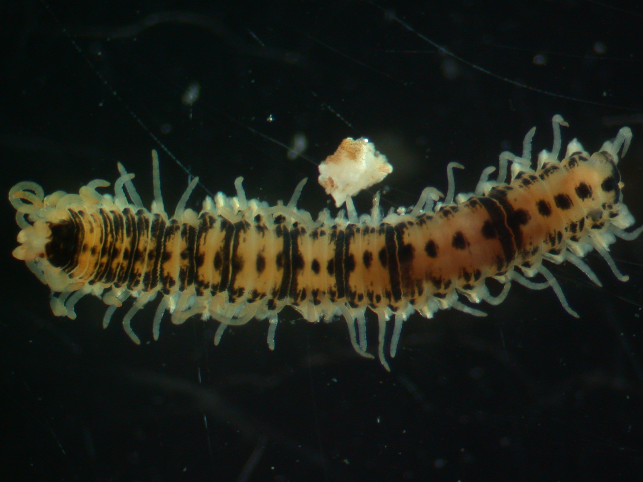
Fig. 1. Syllidae sp. (ICHU22090154), dorsal view.
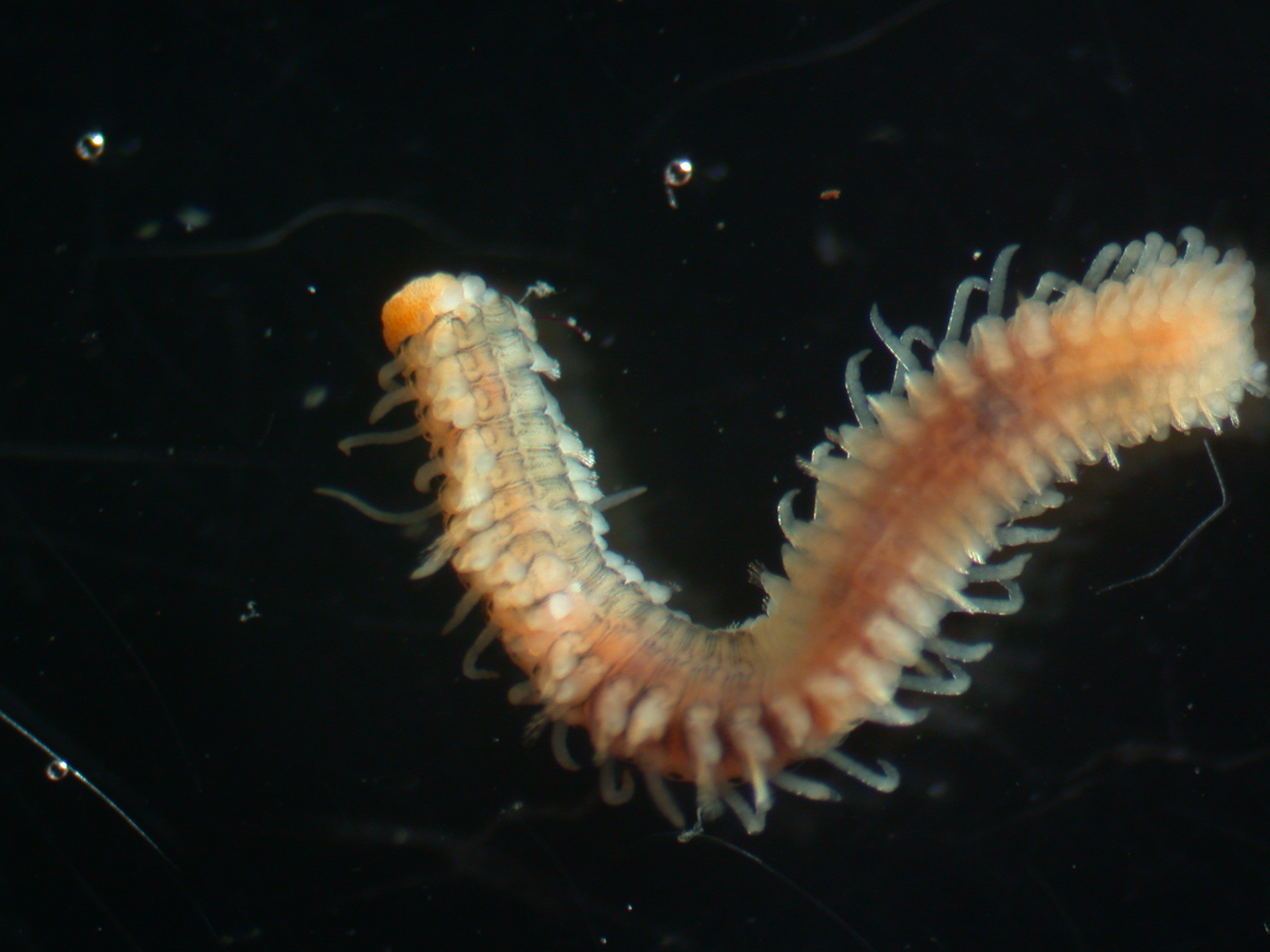
Fig. 2. Syllidae sp. (ICHU22090154), ventral view.
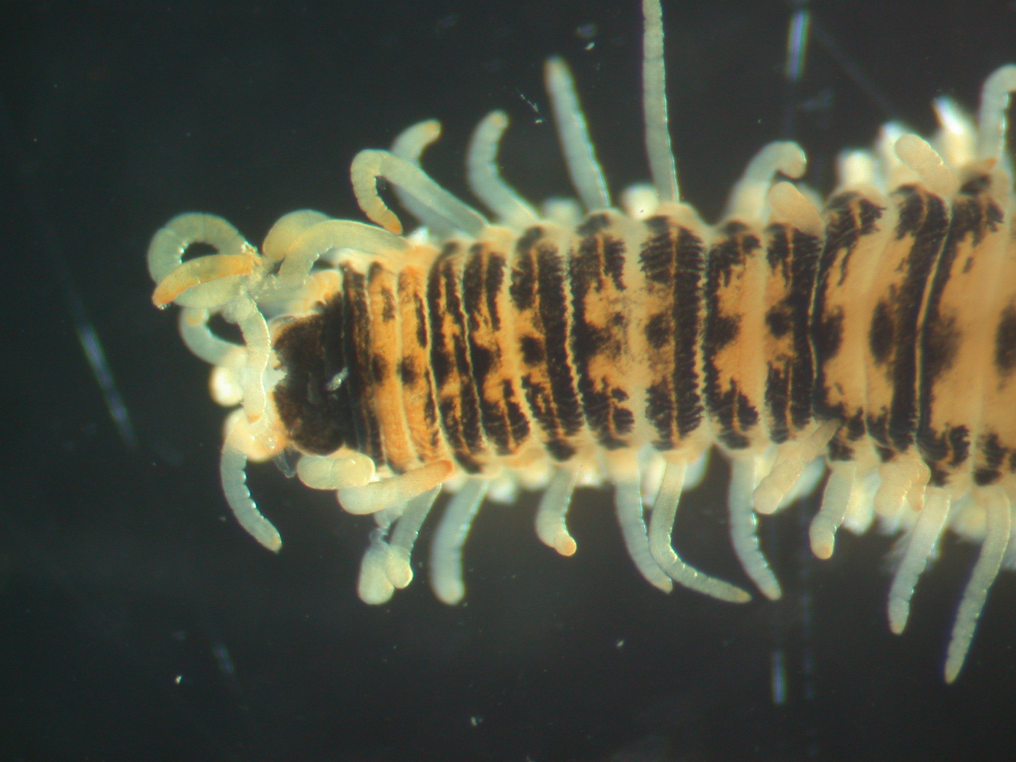
Fig. 3. Syllidae sp. (ICHU22090154), magnification of head.
References
Boom, R., Sol, C. J. A., Salimans, M. M. M., Jansen, C. L., Wertheim-van Dillen, P. M. E., and van der Noordaa, J. 1990. Rapid and simple method for purification of nucleic acids. Journal of Clinical Microbiology 28: 495–503.
Folmer, O., Black, M., Hoeh, W., Lutz, R. and Vrijenhoek, R. 1994. DNA primers for amplification of mitochondrial cytochrome c oxidase subunit I from diverse metazoan invertebrates. Molecular Marine Biology and Biotechnology 3: 294–299.
Tamura, K.,Dudley, J., Nei, M. and Kumar, S. 2007. MEGA4: Molecullar Evolutionary Genetics Analysis (MEGA) software version 4.0. Molecular Phylogenetics and Evolution 24: 1596–1599.