A sea slater was collected on the rocky intertidal zone in Oshoro Bay, Hokkaido, Japan, about 43°12′N, 140°51′E, on 12 June 2013 by Naoya Takeda, then photographed with a Nikon COOLPIX 995 digital camera and fixed in 99% EtOH by Hiroshi Kajihara; the specimen was later identified by HK as Ligia cinerascens Budde-Lund, 1885 by a reference to Nunomura (1995). DNA was extracted from two legs using the silica method (Boom et al. 1990) with some modifications. Extracted DNA was dissolved in 30 µl of deionized water and has been preserved at –20°C. Remaining morphological voucher specimen has been deposited at the Hokkaido University Museum under the catalogue number ICHU2112058 (contact: Dr. Hiroshi Kajihara, kazi@mail.sci.hokudai.ac.jp).
An about 600-bp fragment of mitochondrial cytochrome c oxidase subunit I gene (COI) was amplified by polymerase chain reaction (PCR) using LCO1490 (5′-GGTCAACAAATCATAAAGATATTGG-3′) and HCO2198 (5′-TAAACTTCAGGGTGACCAAAAAATCA-3′) (Folmer et al. 1994). A hot start PCR was performed by a thermal cycler, 2720 Thermal Cycler (Applied Biosystems), in a 20-µl reaction volume containing 1 µl of template total DNA (approximately 10–100 ng) and 19 µl of premix made with 632-µl deionized water, 80-µl Ex Taq Buffer (TaKara Bio), 64-µl dNTP (each 25 mM), 8-µl each primer (each 10 µM), and 0.1-µl TaKara Ex Taq (5 U/µl,TaKara Bio). Thermal cycling condition comprised an initial denaturation at 95°C for 30 sec; 30 cycles of denaturation at 95°C for 30 sec, annealing at 45°C for 30 sec, and elongation at 72°C for 45 sec; and a final elongation at 72°C for 7 min.
The PCR product was purified with the silica method (Boom et al. 1990). Both strands were sequenced with a BigDye® Terminator v3.1 Cycle Sequencing Kit (Applied Biosystems) following the manufacturer's protocol, using the same primer set as the initial PCR amplification. Sequencing was performed with ABI Prism 3730 DNA Analyzer (Applied Biosystems). Chromatogram and sequence data were operated with MEGA v.5 software (Tamura et al. 2011).
Results
Phylum Arthropoda
Subphylum Crustacea
Class Malacostraca
Order Isopoda
Family Ligiidae Leach, 1814
Genus Ligia Fabricius, 1798
Ligia cinerascens Budde-Lund, 1885
[Japanese name: kita-huna-mushi]
(Figs 1, 2)
A total of 559 bp of COI sequence was determined from Ligia cinerascens (see Appendix). However, a nucleotide BLAST search with our sequence at the U.S. National Center for Biotechnology Information resulted in that it was most similar to JX838397 (query coverage 100%; E value 4e-130; identity 82%), which is a sequence from an unidentified acariform mite in the genus Nanorchestes (Young et al. 2012). This suggests that our sequence is likely to have derived from mites associated with the sea slater. Takaku (2000) described the parasitiform mite Thinoseius setifer Takaku, 2000 that was found on the pleopod of Ligia cinerascens from Oshoro Bay. Our sequence may represent either T. setifer or other mites associated with the sea slater, possibly as adults trapped in the leg joint or as eggs laid on the legs.
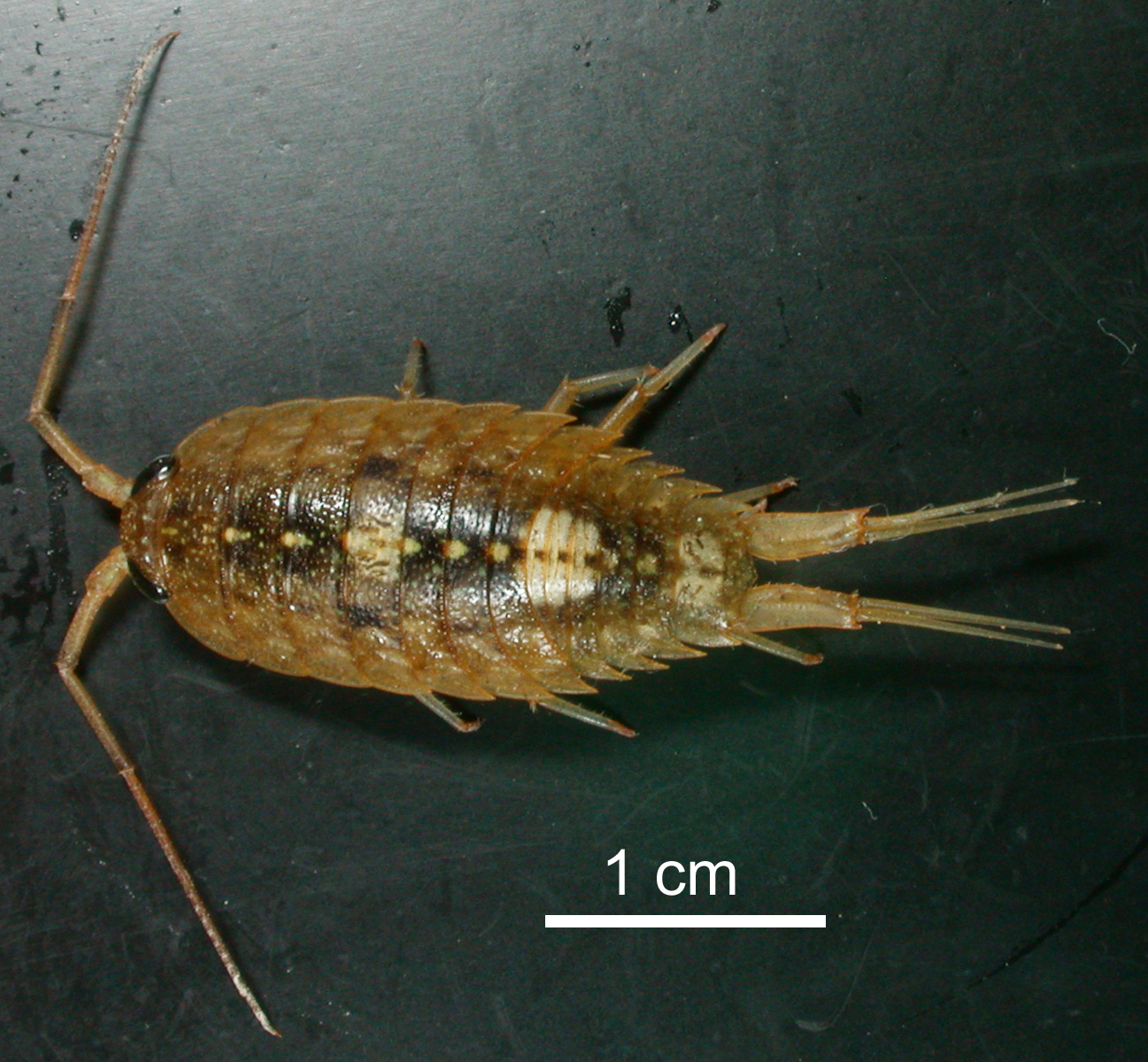
Fig. 1. Ligia cinerascens (ICHU2112058) from Oshoro Bay, dorsal view; the animal had been fixed in EtOH.
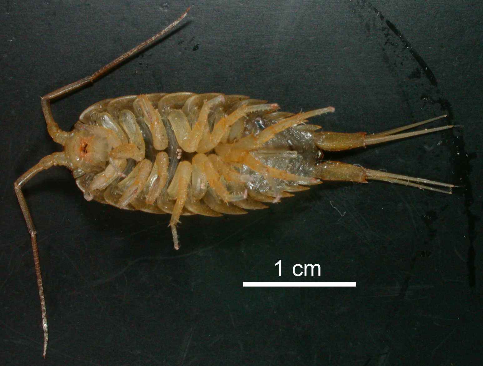
Fig. 2. Ligia cinerascnes (ICHU2112058), ventral view; the pinckish-white 'dots' (three on the sternite of the fourth thoracic segment and two on the merus of the right [bottom on the picture] fifth pereiopod) might represent Thinoseius setifer Takaku, 2000, from which our COI sequence possibly derives.
References
Boom, R., Sol, C. J. A., Salimans, M. M. M., Jansen, C. L., Wertheim-van Dillen, P. M. E., and Van der Noordaa, J. 1990. Rapid and simple method for purification of nucleic acids. Journal of Clinical Microbiology 28: 495–503.
Folmer, O., Black, M., Hoeh, W., Lutz, R. and Vrijenhoek, R. 1994. DNA primers for amplification of mitochondrial cytochrome c oxidase subunit I from diverse metazoan invertebrates. Molecular Marine Biology and Biotechnology 3: 294–299.
Nunomura, N. 1995. Isopoda. Pp. 205–233. In: Nishimura, S. (Ed.) guide to Seashore Animals of Japan with Color Pictures and Keys, Vol. II. Hoikusha, Osaka.
Takaku, G. 2000. Two new mite species of the genus Thinoseius (Acari: Gamasida: Eviphididae) from Japan. Species Diversity 5: 361–374.
Tamura, K., Peterson, D., Peterson, N., Stecher, G., Nei, M., and Kumar, S. 2011. MEGA5: molecullar evolutionary genetics analysis using miximum likelihood, evolutionary distance, and maximum parsimony methods. Molecular Biology and Evolution 28: 2731–2739.
AppendixCOI sequence from ICHU2112058 identidied as Ligia cinerascens Budde-Lund, 1885 from Oshoro Bay, Hokkaido, northern Japan.
AAAAAACTAGTATTAAAGTTTCGATCAGTTAAAAGCATAGTAATAGCCCCAGCCAAAACAGGAAGAGATAATAACAAAAGAAAAGCCGTTACAAAAACAGACCAAACAAAAAGAGGAACTTTTTCAAAGCTTATTCCTACCGCCCGCATATTAATTACAGTAGTAATAAAATTAATAGCACCTAAAATAGAAGAAACACCAGCTAAATGTAAAGAAAAAATAGTAAAATCTACAGAAGTAGTAGAATGGCCTAAAAAGGAAGACAGAGGGGGATAAACAGTTCAACCTGTTCCACTACCAACACCCAAAGAAGAAAAAACTAATATTAACATAGAAGGAGGCAAAAGCCAAAAACTTATATTATTCATACGAGGAAAGGCTATATCAGGAGCTCCAATTATCATTGGAACTAATCAATTTCCAAATCCACCAATTATAATAGGTATCACAGCAAAAAAAATTATAACAAAGGCATGAGAAGTAACATAAACATTATATATATGATCTCTCTCAATAAAACTTCCGGGCTGATTTAACTCTAAACGAACCAAAAAACTCAAAGAAGAACC